Wechat QR code
護(hù)儀廠家,腦電圖機(jī),美倫,美倫醫(yī)療電子有限公司")
TEL:400-654-1200
TEL:400-654-1200

Criteria and technical specifications for brain death
In 1968, the Harvard Medical Committee presented the concept and standard of brain death. Since then, many countries in the world have supported and adopted this standard. In 1970s, China began the theoretical research and clinical practice of brain death determination. In 2003, the Chinese Medical Journal and other major medical journals published the "Brain Death Criteria (Adults) (Draft for Consultation)" and "Technical Criteria for Brain Death Criteria (Adults) (Draft for Consultation)" formulated by the Drafting Group of Brain Death Criteria of the Ministry of Health. In March 2012, the National Health and Family Planning Commission (formerly the Ministry of Health) approved Xuanwu Hospital of Capital Medical University as the National Health and Family Planning Commission brain injury quality control evaluation center. In 2013, on the basis of 10 years'clinical practice and Research on brain death determination, the center revised and improved the above two documents. It is hoped that the new "Standards and Technical Specifications for Brain Death Determination (Adult Quality Control Version)" will be used as a medical industry standard to promote the orderly and standardized development of brain death determination in China. Electroencephalograph
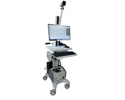
Prerequisites for EEG determination
(1) the reason for coma is clear.
(two) the reversible coma was eliminated for various reasons.
Clinical judgment
(1) deep coma
(two) brain stem reflex disappeared.
(three) without spontaneous breathing, ventilator maintained ventilation, and spontaneous breathing provocation confirmed no spontaneous breathing.
The above 3 clinical judgments must be fully available.Meilun
Confirmatory test
(1) short latency somatosensory evoked potentials (short-1atency)
Somatosensory evoked potential (SLSEP) SLSEP in the median nerve showed bilateral presence of N9 and/or N13 and disappearance of P14, N18 and N20.
Recording technology:
(1) electrode placement: reference electroencephalography international 10-20 system, placing disc electrode or disposable needle electrode:
C'3 and C'4: C'3 and C'4 located in the international 10-20 system and 2 cm after C4 respectively. C'3 or C'4 were called C'4 when stimulating the opposite side.
Fz and FPz: Fz are located at the midpoint of the international 10-20 system and FPz is located at the midpoint of the international 10-20 system.
Cv6: located in the spinous process of the sixth cervical vertebra.
CLi and CLc: 1cm on the ipsilateral or contralateral clavicle midpoint respectively.
(2) electrode lead combination (recording electrode reference electrode): at least 4 channels:
First channel: CLi-CLc (N9);
Second channel: Cv6- Fz, Cv6-FPz or Cv6-CLc (N13);
Third channel: C 'c-CLc (P14, N18);
The fourth channel is C 'c-Fz or C' C -FPz (N20);
(3) electrode impedance: recording, reference electrode impedance less than 5 K ohm;
(4) ground wire placement and impedance: 5 cm above the stimulation point, impedance less than 7K ohm;
(5) analysis time: 50 ms, 100 ms if necessary;
(6) Bandpass: 10~2000 Hz;Meilun
(7) the average number of times: 500~1000 times.
Results: Bilateral N9 and (or) N13 were present, and when P14, N18 and N20 disappeared, they met the criteria of SLSEP brain death.
(two) electroencephalogram showed electrical rest.Meilun

Parameter settings:
(1) Place at least eight recording electrodes according to the international 10-20 system: frontal Fpl, Fp2, central C3, C4, occipital 0l, 02, mesotemporal T3, T4. The reference electrode is located in the double lobe or double mastoid. The grounding electrode is located at the center of the frontal pole (FPz). The common reference electrode is located at the central midline point (Cz).
(2) the impedance between electrode scalp is less than 10k ohm, > 100 ohm, and the impedance of both electrodes should be basically matched.
(3) high frequency filtering 30 to 75Hz, low frequency filtering 0.5Hz or time constant 0.3 s;
(4) sensitivity is 2 V/mm.
Results: When EEG was resting (EEG activity < 2 mu V), EEG met the criteria of brain death.
Electroencephalograph
(3) Transcranial Doppler ultrasound (TCD) showed that the blood flow of anterior and posterior intracranial circulation showed oscillatory waves, small systolic waves or disappearance of blood flow signals.
Parameter setting: electroencephalograph
(1) set the appropriate output power;
(2) set the sampling volume: 10~15 mm;
(3) adjust gain: adjust the gain strength according to the sharpness of spectrum display;
(4) adjust the speed scale: make the spectrum displayed on the screen in proper size.
(5) adjust the baseline: the upper and lower frequency spectrum is displayed on the screen.
(6) adjust SNR: clearly display spectrum and minimize noise;
(7) screen scanning speed: 6 to 8s per screen;
(8) set Doppler frequency filtering: low filtering state (less than 50 Hz).
Inspection site:
(1) Temporal window: In supine position, the middle cerebral artery (MCA) was detected in the area of horizontal connection between eyebrow arch and auricular margin.
(2) occipital window or paraoccipital window: the vertebral artery and basilar artery were detected in supine position (raising the head) or lateral position, beside the foramen magnum of occipital bone or foramen magnum of occipital bone under the occipital trochanter.
(3) eye window: supine position, on the closed upper eyelid, the contralateral side.
MCA and ipsilateral internal carotid artery siphon (internal carotid artery siphon) segments.
The results were as follows:
(1) Judging vessels: bilateral MCA was the main judging vessel in the anterior circulation, and basilar artery was the main judging vessel in the posterior circulation.
(2) determine blood flow spectrum:
(1) reverberating flow: systolic and diastolic reverse flow signals were observed in one cardiac cycle, direction of flow index (DFI) < 0.8, DFI = 1-R / F (R: reverse flow velocity, F: positive flow velocity).
(2) Early systolic small systolic peaks in early systole: unidirectional positive blood flow signal in early systole, duration less than 200 ms, flow rate less than 50 cm/s;
3. The blood flow signal disappeared.
(3) the number of determination: interval 30 min, 2 times.
Results: Both anterior and posterior intracranial circulation were above any blood flow spectrum, which accorded with TCD criteria for brain death.
(four) confirm the order of trials.
The optimal sequence of the confirmatory test was SLSEP, EEG and TCD. Confirm that at least 2 items should meet the criteria of brain death.
Decision step
Brain death judgment is divided into the following three steps: step 1 clinical judgment of brain death, in accordance with the judgment criteria (deep coma, brainstem reflex disappearance, no spontaneous breathing) can enter the next step. The second step of the brain death confirmation test, at least 2 items meet the criteria for brain death can enter the next step. In the third step, the spontaneous breathing test of brain death was conducted to verify the spontaneous breathing. These 3 steps are consistent with the criteria for determining brain death and are confirmed as brain death.
Judge
There are at least two doctors who perform brain death judgment, and they are required to be medical practitioners who have been engaged in clinical work for more than five years.